If you live in or around Perth, Ontario and you’ve been told you need shockwave therapy — or you’ve read that it can help stubborn heel or tendon pain — you may have found it’s hard to find nearby. Shockwave isn’t offered at most local clinics. The good news: Nobility Chiropractic & Wellness in Smiths Falls is only about a 20-minute drive from Perth, and shockwave therapy is one of our specialties.
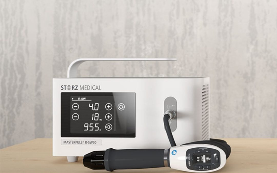
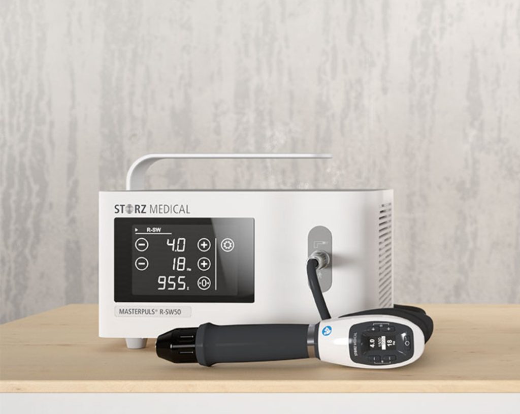
Our Swiss-engineered Storz Masterpuls R-SW50 radial shockwave unit.
What Is Shockwave Therapy?
Shockwave therapy (radial extracorporeal shockwave therapy, or ESWT) is a non-invasive treatment that delivers acoustic pressure waves into injured tissue. Those waves boost blood flow, break down scar tissue and calcifications, and kick-start your body’s natural repair process — no needles, no medication, and no downtime. You can read the full details on our shockwave therapy page.
Does It Actually Work?
For the right conditions, yes — and it’s well studied. Multiple systematic reviews have found shockwave therapy to be an effective, safe treatment for chronic plantar fasciitis (persistent heel pain), with high success rates reported and a low risk of side effects.12 It works by boosting blood flow and stimulating your body’s natural repair response — no needles, no medication, and no downtime — which makes it a strong option for stubborn heel and tendon problems that haven’t responded to other care.
Because shockwave therapy isn’t widely available in the immediate Perth area, we’re often the closest option — and for a targeted, specialized treatment like this, a 20-minute drive is well worth it. Sessions take just 10–15 minutes, most people notice meaningful improvement within 3 to 5 treatments, and we treat patients from Perth, Carleton Place, and across Lanark County every week.
Plantar fasciitis is one of the most common heel-pain complaints I see at the clinic. The classic version sounds like this: sharp pain on the bottom of the heel with the first few steps in the morning, eases after walking around for a few minutes, then returns after long sitting or at the end of a long day on your feet. Sometimes it’s been going on for weeks. Sometimes it’s been going on for over a year and you’ve started compensating without realizing it.
This post explains what plantar fasciitis actually is, how I assess it, what treatment looks like at the clinic, and what you can reasonably expect from the recovery process.
A note before we get into it: this is general information about how I approach plantar fasciitis clinically, not specific medical advice for you. Heel pain has multiple possible causes — actual diagnosis requires assessment in person.
What plantar fasciitis actually is
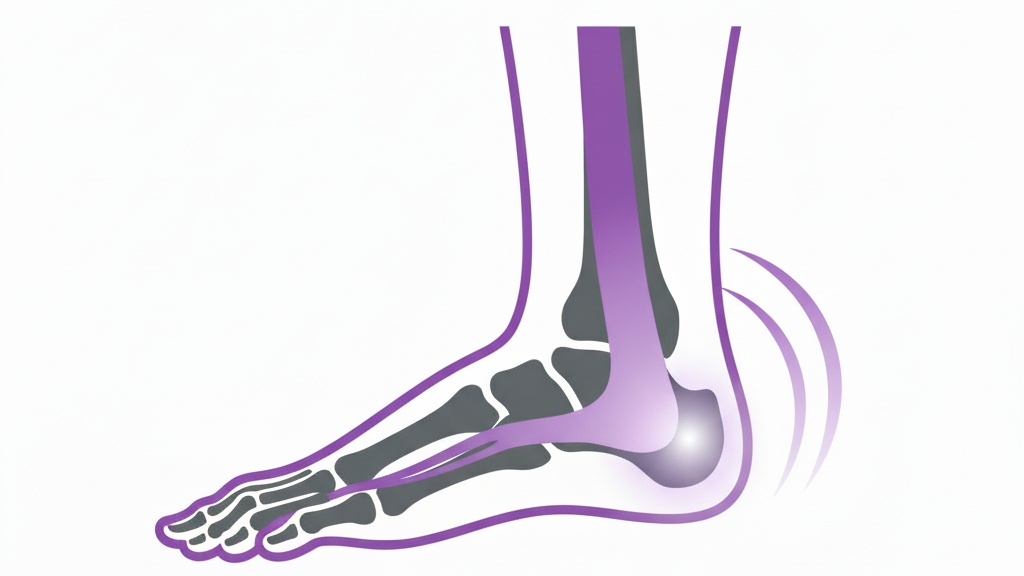
The plantar fascia is a thick band of connective tissue that runs along the bottom of your foot, from the heel bone forward to the base of your toes. Its job is to support the arch and absorb load with every step.
“Plantar fasciitis” technically refers to inflammation of that tissue, but the more accurate term in many cases is plantar fasciopathy — chronic degeneration of the fascia rather than acute inflammation. Histological studies of long-standing cases show that the tissue often has the structural changes of chronic overload, not the cellular signs of active inflammation. Practically, this matters because anti-inflammatory approaches alone tend not to fix the underlying mechanical problem driving the pain.
The pain itself usually shows up at the medial heel — the inside of the bottom of your heel where the fascia attaches to the heel bone. That’s the spot that hurts when you press on it, and that’s where the morning-step pain originates.
Why the foot isn’t always the problem
One of the things I look for early in any plantar fasciitis assessment is whether the foot is the actual driver of the problem, or whether it’s the place that’s paying the price for dysfunction further up the chain.
The foot is the end of a long kinetic chain. Restricted ankle dorsiflexion, tight calves, hip mobility limitations, glute weakness, and even old low back issues can all change how load travels through the lower extremity with every step. Walk 10,000 steps a day with a slightly off-pattern landing, and the fascia ends up absorbing more than it’s designed for. That’s the version of plantar fasciitis that doesn’t respond to “ice and stretch the foot” — because the foot isn’t where the problem started.
So the assessment looks at:
Local foot and ankle mobility, particularly ankle dorsiflexion
Calf and Achilles tightness
Hip and glute function on both sides
Pelvic position and lumbar mobility
Walking pattern and gait mechanics
Footwear, training surface, and recent changes in activity
Sometimes the foot is the whole story. Often it isn’t.
How I treat plantar fasciitis at the clinic
Treatment is always individualized to what the assessment actually shows, but the typical components are:
Manual therapy to the foot and ankle. This includes joint mobilization of restricted tarsal and ankle joints, soft tissue work to the plantar fascia itself, calf muscles, and any associated trigger points. Manual therapy and stretching for plantar heel pain has reasonable supporting evidence, particularly for soft tissue mobilization techniques.[1]
Addressing dysfunction up the chain. If the assessment shows hip, pelvic, or lumbar contributions to the problem, those get addressed too. Adjusting only the foot and ignoring the rest of the chain tends to produce short-term relief without lasting change.
Loading and stretching protocols. Plantar fascia-specific stretching, calf flexibility work, and progressive loading exercises are core parts of recovery. The current evidence supports stretching, heel raises, and graded loading as foundational components of conservative treatment.[2] The specifics depend on what your foot needs and how irritable it currently is.
Footwear and activity guidance. Sometimes the most important intervention is changing what you’re doing during the other 23 hours of the day — different shoes for work, a different surface for your runs, an activity-modification plan that lets the tissue actually heal between loading sessions.
Shockwave therapy when indicated. Extracorporeal shockwave therapy (ESWT) has the strongest evidence base of any single intervention for chronic plantar fasciitis, with high-quality systematic reviews showing meaningful effects on both pain and function.[3] Shockwave isn’t always the first thing we use — but for cases that have been going on for a while or aren’t responding to manual therapy and loading, it’s a useful tool. (Shockwave is currently available at our Stittsville clinic, Nobility Performance, with plans to bring it to Smiths Falls as well.)
What recovery actually looks like
Realistic expectations matter here, because plantar fasciitis recovery isn’t always linear and the timeline depends a lot on how long it’s been going on.
Early stages (first few weeks): Morning pain often improves first. Sharp first-step pain typically eases within 2-4 weeks of consistent treatment plus home work. This is encouraging but it isn’t the finish line — the tissue is still vulnerable, and stopping treatment at this point is the most common reason it comes back.
Middle stages (4-12 weeks): The fascia and surrounding structures are loading better. End-of-day soreness fades. We’re typically working on returning to fuller activity, addressing whatever drove the original overload, and progressing your loading program.
Longer-term (3-6 months for chronic cases): Stubborn cases that have been going on for a year or more sometimes take this long to fully resolve. The fascia regenerates slowly. The good news: even chronic cases generally do respond to conservative treatment given enough time and the right inputs.
Most plantar fasciitis cases resolve with conservative care over 3-6 months. A small percentage don’t and require escalation — surgical consultation is sometimes part of that conversation, but it’s a last resort, not a first option.
What you can do at home
Some of the highest-leverage things you can do between visits:
Calf stretching, multiple times per day. Tight calves are a major contributor to plantar fascia overload. Both straight-leg and bent-knee versions cover the gastrocnemius and soleus.
Plantar fascia-specific stretching. Pulling your toes back toward your shin, especially first thing in the morning before you stand up, can dramatically reduce that first-step pain.
Roll the bottom of the foot. A frozen water bottle or a lacrosse ball under the arch for a few minutes a day. Combines tissue mobilization with cold for soreness.
Manage load. If running or walking volume has crept up recently, dial it back temporarily. The fascia needs time to recover between loading sessions.
Avoid going barefoot on hard surfaces while symptoms are active. Supportive footwear in the morning before you stand up makes a real difference for some people.
Show up for your sessions and do your homework. The clinical work and the home work compound each other. Doing only one cuts the result roughly in half.
When to come in
A few situations where booking an assessment is the right move:
Heel pain that’s been around for more than 2-3 weeks and isn’t improving
Morning first-step pain that’s affecting your daily routine
Pain that’s started limiting your training, walking, or work
An old plantar fasciitis episode that keeps coming back
Heel pain that doesn’t quite fit the typical pattern (worth ruling out other causes)
Not all heel pain is plantar fasciitis. Stress fractures of the heel bone, fat pad atrophy, nerve entrapments, Achilles tendon issues, and a few other conditions can all cause heel pain. Part of the value of a proper assessment is making sure we’re treating the right thing.
Common questions
How quickly will I notice a difference? For most people, morning pain starts easing within 2-4 weeks. Full recovery from a recent case is typically 6-12 weeks. Long-standing cases take longer — sometimes 3-6 months. Improvement isn’t always linear; some weeks you’ll feel a clear change, others will feel stuck.
Do I need imaging? For a typical presentation, no. Plantar fasciitis is diagnosed clinically — history and physical exam are sufficient in most cases. Imaging becomes relevant if the presentation is atypical, the pain isn’t responding to conservative care, or there’s reason to suspect a stress fracture or other diagnosis.
Can I keep training? Usually yes, with modifications. Total rest isn’t typically the answer — the fascia responds well to graded loading. The plan we’d build for an active patient usually involves dialing back the most aggravating activities while maintaining conditioning through movements that don’t load the foot the same way (cycling, swimming, strength work that isn’t standing-heavy).
What about orthotics? Sometimes useful, especially during the active recovery phase. Off-the-shelf supportive insoles work for many people. Custom orthotics make sense for specific situations but aren’t always necessary. We figure out what’s right based on the assessment, not based on a default protocol.
Will it come back after I’m better? It can, particularly if the underlying contributing factors aren’t addressed. The work we do during recovery on calf flexibility, hip and glute function, footwear, and load management is also what reduces the chance of recurrence.
What about Applied Kinesiology for this? AK assessment is part of how I work and is particularly useful here for identifying inhibition patterns in the foot intrinsics, calves, and hip stabilizers that contribute to fascial overload. (For more on how AK fits into clinical assessment, see our post on Applied Kinesiology.)
Booking an assessment
If you’re dealing with heel pain in Smiths Falls, Lanark County, or the surrounding region, a thorough assessment is the right starting point. We’ll figure out what’s actually driving the pain, build a treatment plan around your specific findings, and give you the homework that supports the clinical work.
Charles R, Fang L, Zhu R, Wang J. The effectiveness of shockwave therapy on patellar tendinopathy, Achilles tendinopathy, and plantar fasciitis: a systematic review and meta-analysis. Front Immunol. 2023;14:1193835. doi.org/10.3389/fimmu.2023.1193835
If you train hard on weekends but grind at a desk during the week, your workstation can either support recovery—or sabotage it. This step‑by‑step setup reduces neck, shoulder, and low‑back strain while keeping you ready for sport.
Low back/hips: Prolonged flexion = hip flexor tightness, inhibited glutes, achy lumbar. Key idea: Your “next posture is your best posture.” We aim for neutral setup + frequent, small position changes.
10‑minute workstation setup checklist
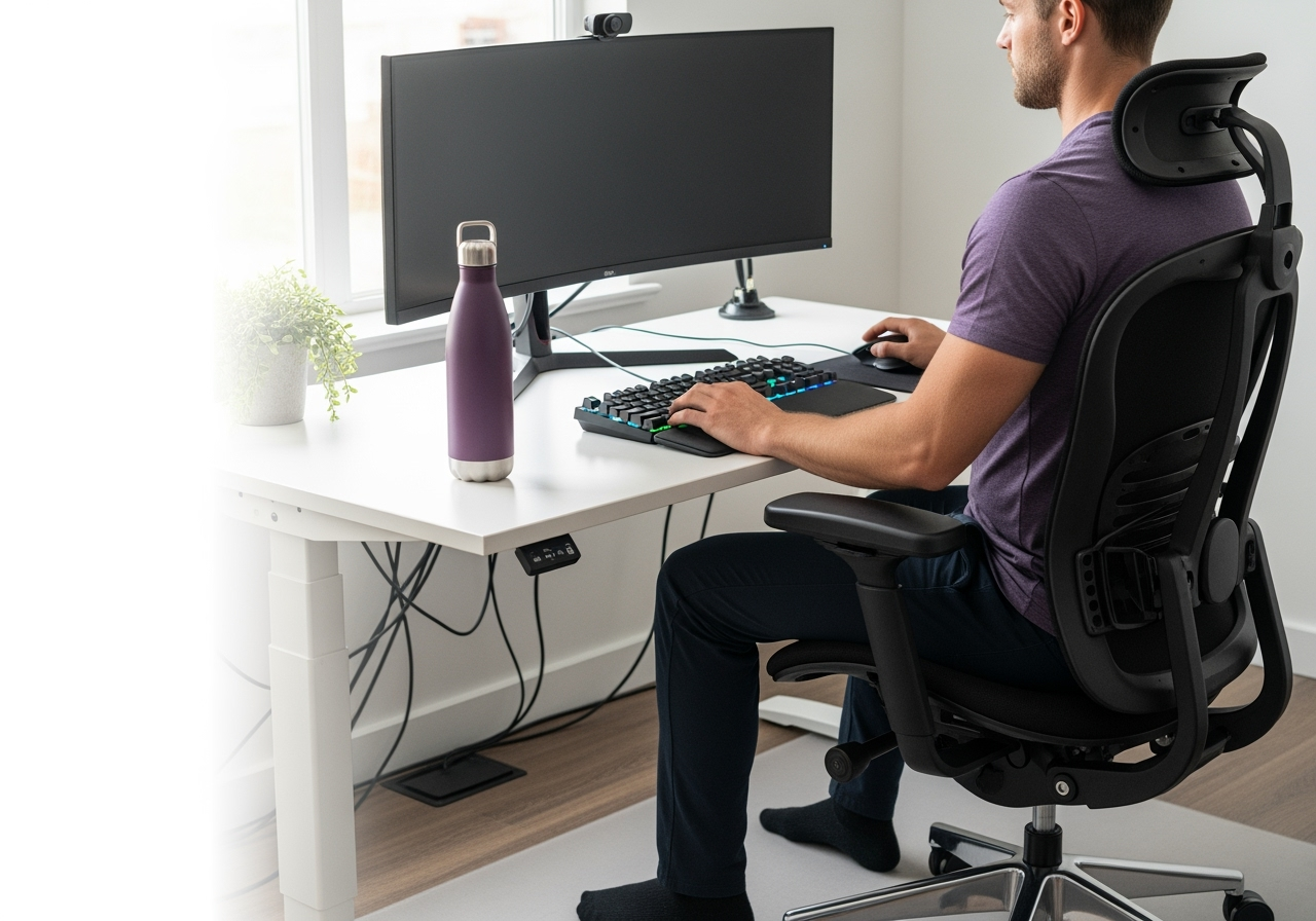
Chair setup
Seat height: Sit so knees and hips are level or hips slightly higher. Feet flat on floor.
Seat depth: 2–3 fingers between the seat edge and the back of your calf.
Lumbar support: Support the natural curve; if none, add a small cushion or rolled towel.
Armrests: Height so shoulders relax (not shrugged). Elbows near 90–110° and close to body. Common fix: If your feet don’t reach the floor, use a footrest or a firm box.
Monitor position
Height: Top of the screen at eye level (or 1–2 inches below if you wear progressive lenses).
Distance: About an arm’s length (50–70 cm). Move closer if you lean forward to read.
Centering: Align the primary monitor with your nose; dual monitors used equally should be centered; if one is primary, center that one and angle the secondary.
Keyboard and mouse
Placement: Close to the body so elbows stay by your sides; forearms parallel to floor.
Keyboard tilt: Flat or slightly negative tilt to avoid wrist extension.
Mouse: Same height/plane as keyboard. Keep wrist neutral—move from shoulder, not just the wrist. Consider a larger mouse if you death‑grip a tiny one.
Desk height
Ideal: Around elbow height when shoulders are relaxed. For most, 25–30 inches; adjust to you, not the spec.
Standing desk: Set the same elbow‑height rule. Keep screen height consistent with sitting.
Laptop users
Use a laptop stand to lift the screen to eye level + external keyboard/mouse. This is non‑negotiable for daily use.
Lighting and glare
Place monitor perpendicular to windows to reduce glare.
Use a task light to keep the screen dimmer than your paper/keyboard.
Cable and reach management
Keep daily‑use items within the forearm “easy reach” zone.
Move the phone/headset to the non‑mouse side to alternate load.
Micro‑breaks and mobility plan (2 minutes per hour)
Timing: 30–60 seconds every 30 minutes; 2 minutes every hour. Set a timer or use software nudges.
Movement snacks:
Chin nods + retractions x10 (small, gentle)
Shoulder blade slides: elbows at sides, squeeze down/back x10
Thoracic extension over chair back x5 breaths
Hip opener: stand, lunge stretch 30s/side
Calf raises x15 or brisk walk to water
Standing rotation drill for golfers/pickleball/hockey: hands across chest, slow trunk rotations x10 each way.
Common mistakes (and easy fixes)
Perching on front of chair → Slide back and use lumbar support.
Shrugged shoulders at the keyboard → Lower armrests/desk; bring keyboard closer.
Monitor too low → Stack books or use a riser; stop craning the neck.
Wrist resting on sharp desk edge → Add a soft desk mat; keep wrists neutral.
All‑day standing → Alternate: sit 30–45 min, stand 15–20 min; change positions often.
Footrest: A stable box works; adjustable footrests add comfort.
FAQ
Q: What is the correct monitor height?
A: Top of screen at eye level; arm’s length away; adjust for progressive lenses.
Q: Is a standing desk better?
A: It’s a change tool, not a cure. Alternate positions and keep elbows at desk height.
Q: What if my feet don’t touch the floor?
A: Use a footrest to keep knees/hips level and reduce low‑back strain.
Q: Do I need an expensive chair?
A: Adjustability matters most. Add a lumbar roll before upgrading.
When to get help If you’re dealing with recurring neck/shoulder headaches, numbness/tingling, or stubborn low‑back pain, a targeted assessment can identify mobility or motor‑control gaps your setup can’t fix alone.
Ready for a personalized ergonomic tune‑up?
Book an assessment with Nobility Chiropractic & Wellness in Smiths Falls. Online booking via Jane, or call/text 343.801.0094.
Whether you’re a competitive athlete, a weekend warrior, or simply someone who likes to stay active, injuries have a way of derailing your momentum. The frustrating part is that most injuries aren’t random — they trace back to a specific weak link: a muscle that isn’t firing properly, a joint that won’t move the way it should, or a left-to-right imbalance you can’t feel until it lets you down. At Nobility Chiropractic & Wellness in Smiths Falls, we use applied kinesiology to find those weak links and address them — so you recover faster and stay in the game longer.
Muscle testing helps pinpoint which muscles aren’t pulling their weight.
What Is Applied Kinesiology?
Applied kinesiology (AK) is a hands-on assessment approach that looks at how your muscles, joints, and nervous system are working together. Rather than treating a sore spot in isolation, we combine manual muscle testing with movement and neurological assessment to map out where your body is compensating — a glute that isn’t switching on, a stiff ankle changing how you land, or a stabiliser that fatigues too early. New to the idea? Start with our primer on what applied kinesiology is and how it helps correct muscle imbalances.
Why Athletes Get Injured — and Where AK Fits In
Athletes ask more of their bodies, and small imbalances add up under load. The research backs this up: left-to-right strength differences and movement asymmetries are consistently linked to a higher risk of injury.1 Put simply, when one side is quietly doing more than its share, it’s often only a matter of time before something gives. Applied kinesiology is built to catch that kind of hidden weak link before it becomes a strain or a tear. For the flip side of the same coin, see the connection between muscle testing and peak athletic performance.
Recover Faster by Treating the Cause, Not Just the Symptom
When you’re already hurt, the fastest route back is fixing what actually caused the problem — not just calming the painful area. By pinpointing the muscles that aren’t pulling their weight and the joints that aren’t moving freely, we can target treatment with adjustments, soft-tissue work, and specific corrective exercises. That means less guesswork and a more direct path back to training. It’s the same root-cause approach we take with common sport injuries like hamstring strains and ACL rehabilitation.
Prevent the Next Injury
The best injury is the one that never happens. Decades of sports-medicine research show that structured, movement-based training can cut injury rates dramatically: a systematic review in the British Journal of Sports Medicine found neuromuscular training reduced injuries in young athletes by roughly a third,2 and programs such as FIFA 11+ have produced similar drops in ankle and lower-limb injuries.3 Applied kinesiology makes that prevention personal — instead of a generic warm-up, we build a corrective plan around your specific weak links. Learn more about how we use it to enhance athletic performance and the broader role of chiropractic in sports performance.
What to Expect at Nobility Chiropractic
Your first visit starts with a thorough assessment — health history, a movement screen, and muscle and joint testing — so we understand how your body is really working. From there, Dr. Bryan Pankow builds a tailored plan that pairs hands-on treatment with targeted exercises, then re-tests to make sure the change sticks. The goal is simple: get you moving well, performing better, and staying healthy.
Book with a Sport-Focused Chiropractor in Smiths Falls
If you’re an athlete in Smiths Falls or Lanark County who’s tired of the same nagging injuries, applied kinesiology can help you find the root cause and build a plan around it. Learn more about applied kinesiology at Nobility, or book an appointment to get started.
Applied Kinesiology (AK) is a biomechanical and neuromuscular assessment framework I’ve trained in for the better part of a decade. Used well, it’s one of the more useful tools I have for figuring out what’s actually going on with an athlete’s body — particularly when conventional assessment hasn’t given clear answers.
The short version: AK uses manual muscle testing to detect inhibition and facilitation patterns — muscles that aren’t firing properly because the nervous system has dialed them down, often in response to old injuries, joint dysfunction, or compensation patterns that have been running for years. Once those patterns are identified, treatment can be targeted at what’s actually driving them rather than at where the pain happens to be felt.
This post explains how I use AK in practice, the underlying neuromuscular concepts the work is built on, and where the evidence sits.
A note before we get into it: this is general information about how I practice, not specific medical advice. Whether AK assessment is useful for your particular situation depends on what you’re dealing with — and that’s a conversation in person, not on the internet.
What “muscle inhibition” actually is
The core concept under AK is that muscles can be neurologically inhibited — not weak from lack of training, not torn or injured, but turned down by the nervous system. The muscle is structurally fine; it’s just not getting the signal to fire properly.
This isn’t a fringe idea. It’s a well-established concept in sports medicine and physiotherapy — particularly under the term arthrogenic muscle inhibition (AMI). The classic example is what happens to the quadriceps after an ACL injury: the muscle is intact, but neural signals from the swollen, painful joint suppress activation. Athletes can lose 30-50% of their quad strength even when the muscle itself is uninjured.[1]
A 2018 scoping review in the British Journal of Sports Medicine identified arthrogenic muscle inhibition as a critical factor limiting recovery after ACL reconstruction, and reviewed evidence-based interventions for addressing it.[2] This is mainstream sports medicine, published in one of the most-cited sports medicine journals in the world.
The same neurological mechanisms — joint inflammation, faulty proprioceptive input, altered reflex patterns, central nervous system adaptation — produce inhibition in less dramatic situations all the time. Old ankle sprains affect glute activation. Chronic low back pain alters core firing patterns. A history of shoulder impingement can dial down rotator cuff recruitment for years after the original injury healed.
None of that shows up on an MRI. None of it shows up on a strength test where you ask someone to push with maximum effort against a hand dynamometer. But it shows up on assessment when you know what you’re looking for — and it directly affects how an athlete moves, performs, and gets injured.
How I use AK in practice
What an AK assessment session actually looks like at the clinic:
1. History and movement screen. Same as any assessment — current complaints, training history, prior injuries, what you’re trying to accomplish.
2. Muscle testing as part of the physical exam. I assess the strength and recruitment quality of muscles relevant to your complaint and your sport. The test is: can this muscle hold against a controlled force in a specific position, with the right timing and quality of activation? I’m not looking for raw strength — I’m looking for inhibition, asymmetry, and recruitment patterns that don’t match what your body should be capable of.
3. Identifying drivers. When I find a muscle that’s inhibited, the next question is “why?” Sometimes the answer is local — a joint restriction, a fascial adhesion, a trigger point in a synergist or antagonist muscle. Sometimes the answer is upstream or downstream — a hip problem creating compensatory patterns at the knee, a thoracic restriction altering shoulder mechanics. AK gives me a real-time framework for testing these hypotheses rather than guessing.
4. Treatment with retest. If I think a joint restriction is driving an inhibition pattern, I’ll address it and retest. If the muscle now activates properly, that’s confirmation. If it doesn’t, I look elsewhere. The test-retest cycle is what separates AK from “I think I know what’s wrong, let me try this and hope for the best.”
5. Homework matched to findings. The exercises and self-care I recommend are based on what the assessment actually showed, not on a generic protocol for your complaint.
What the literature supports — and where it’s mixed
Worth being honest about the evidence picture:
The mechanism of muscle inhibition is well-established. AMI is a real, measurable, neurologically-mediated phenomenon. The literature is clear that joint pathology, inflammation, and altered afferent input can produce profound inhibition of surrounding musculature, and that interventions aimed at restoring normal joint function and afferent input can help reverse it.[1][2]
Spinal manipulation produces measurable changes in muscle activation. A 2018 RCT in elite athletes showed significant increases in maximum voluntary contraction and corticospinal excitability after a single manipulation, persisting 30-60 minutes.[3] A separate study found 44-54% increases in motor evoked potentials following manipulation, suggesting changes happen at the level of cortical drive — exactly the kind of effect AK practitioners describe clinically.[4] When AK assessment leads me to deliver an adjustment and I see immediate facilitation of a previously inhibited muscle, the underlying neuromechanism is supported.
Manual muscle testing reliability is the honest weak link. A 2020 study examining force profiles of testers performing manual muscle tests found significant differences between experienced and beginner testers, and even experienced testers showed inter-tester variability and partial intra-tester reproducibility issues.[5] Older research on traditional manual muscle testing in clinical populations also showed reproducibility problems compared to instrumented testing.[6]
The honest interpretation: MMT is a tester-skill-dependent tool. Done by someone with significant training and standardization, it produces consistent results. Done by someone untrained or inconsistent in their force application, it doesn’t.
This is part of why I’ve invested 400+ hours of additional training beyond the initial ICAK certification — including multiple 100-hour AK course series, clinical pearls work with the late Dr. Wally Schmitt, and instruction with Dr. David Leaf at ICAK international meetings. The certification covers the basics; proficiency comes from deliberate practice, study with senior practitioners, and consistent recalibration. It’s also why MMT is best used as one component of a comprehensive assessment, not the sole basis for diagnostic conclusions.
Where AK earns its keep
The honest version of where this approach pays off:
Identifying neuromuscular inhibition patterns that don’t show up on standard strength testing or imaging
Real-time test-retest assessment to confirm whether an intervention actually worked
Untangling complex compensation patterns where pain in one location is being driven by dysfunction elsewhere
Building an individualized treatment and homework plan based on what your specific body needs, rather than a protocol
Performance work with athletes whose strength on paper doesn’t match how they actually move and generate force in their sport
For anything outside the musculoskeletal scope — bloodwork, imaging, allergy testing, nutritional analysis, or specialist medical care — the right answer is referral to the appropriate professional. I work within my scope.
Why this approach matters for athletes
Most assessment systems for athletes rely on either subjective complaint (“my hamstring feels tight”) or maximum-effort strength testing (“can you push as hard as possible against this”). Both have value, but both miss the layer where most performance-limiting dysfunction actually lives.
The athletes I see most often aren’t catastrophically injured. They’re partially inhibited. The hamstring that’s “tight” is often actually under-recruited. The shoulder that “feels off” has a serratus anterior that’s not firing properly. The knee that “doesn’t track right” has a glute that’s not doing its job.
You can train through these patterns for a long time before they become an injury — but they cost you in performance, and they raise your injury risk over time. AK gives me a way to find them earlier and address them more precisely.
Common questions
What does an AK assessment add to my visit? It adds a structured, testable framework for figuring out what your specific body needs on the day you come in. Muscle testing gives me real-time information that informs which interventions to use, in what order, and whether they actually worked before you walk out the door. The treatment itself — adjustments, soft tissue work, exercise prescription — is similar to what you’d see in any sport-focused chiropractic visit; the assessment is what makes the plan more individualized.
Do I need to “believe in” AK for it to work? No. AK isn’t a faith-based system in how I practice it. The muscle either tests strong or it doesn’t. The intervention either changes the test or it doesn’t. You can be skeptical of the framework and still benefit from it, the same way you can be skeptical of “core stability training” and still benefit from learning to brace properly.
Is this covered by insurance? The chiropractic care itself is. There’s no separate “AK fee” — assessment is part of how I work, not an add-on service.
How long does an AK-informed visit take? About the same as a regular chiropractic appointment. Initial assessments run longer (45-60 minutes) because of the comprehensive movement and muscle screen. Follow-ups are shorter (15-30 minutes) since we’re working from a known baseline.
Do you do AK on non-athletes? Yes. The framework is just as useful for desk workers with chronic pain, post-surgical patients managing residual deficits, or anyone whose body has compensation patterns from old injuries. It’s not athlete-exclusive — that’s just where the gains are most visible.
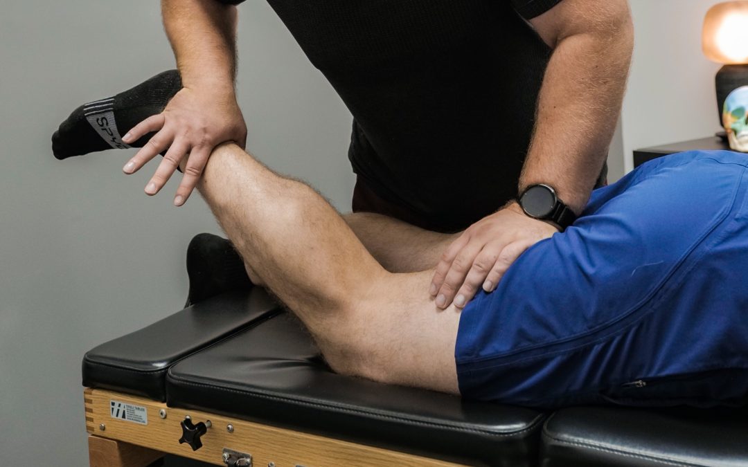
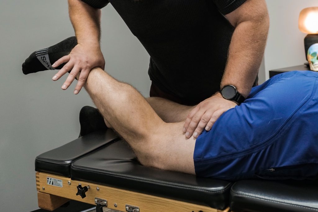
What does an AK assessment actually feel like? Mostly like a thorough orthopedic exam. I’ll have you in different positions — supine, prone, standing, sometimes mid-movement — and apply controlled force to various muscles while you resist. It’s not painful, it’s not strenuous, and you don’t need to be at maximum effort. The goal is to feel how your body responds, not how hard you can push.
If you want to try it
I’m an ICAK-certified Applied Kinesiology practitioner with 400+ hours of additional AK training beyond the certification, including study with the late Drs. Wally Schmitt and David Leaf. I see athletes, tactical professionals, and active people across both Nobility Chiropractic & Wellness in Smiths Falls and Nobility Performance in Stittsville.
If you’re an athlete dealing with persistent issues that haven’t responded to conventional approaches, or you want a more individualized assessment than a generic protocol-based visit, this is the work.
Pietrosimone B, Lepley AS, Kuenze C, et al. Arthrogenic Muscle Inhibition Following Anterior Cruciate Ligament Injury. J Sport Rehabil. 2022;31(6):694-706. doi.org/10.1123/jsr.2021-0128
Sonnery-Cottet B, Saithna A, Quelard B, et al. Arthrogenic muscle inhibition after ACL reconstruction: a scoping review of the efficacy of interventions. Br J Sports Med. 2019;53(5):289-298. doi.org/10.1136/bjsports-2017-098401
Christiansen TL, Niazi IK, Holt K, et al. The effects of a single session of spinal manipulation on strength and cortical drive in athletes. Eur J Appl Physiol. 2018;118(4):737-749. doi.org/10.1007/s00421-018-3799-x
Haavik H, Niazi IK, Jochumsen M, et al. Impact of Spinal Manipulation on Cortical Drive to Upper and Lower Limb Muscles. Brain Sci. 2017;7(1):2. doi.org/10.3390/brainsci7010002
Bittmann FN, Dech S, Aehle M, Schaefer LV. Manual Muscle Testing—Force Profiles and Their Reproducibility. Diagnostics (Basel). 2020;10(12):996. doi.org/10.3390/diagnostics10120996
Escolar DM, Henricson EK, Mayhew J, et al. Clinical evaluator reliability for quantitative and manual muscle testing measures of strength in children. Muscle Nerve. 2001;24(6):787-93. doi.org/10.1002/mus.1070